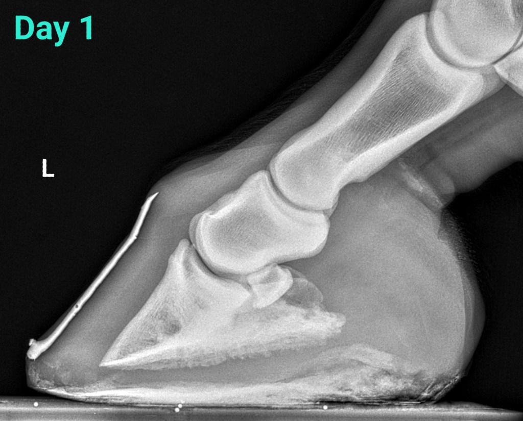
Acute, mild laminitis episode
28 year old Haflinger gelding – overweight. He was placed in NANRIC Ultimates. Caloric intake was reduced and exercise was restricted.

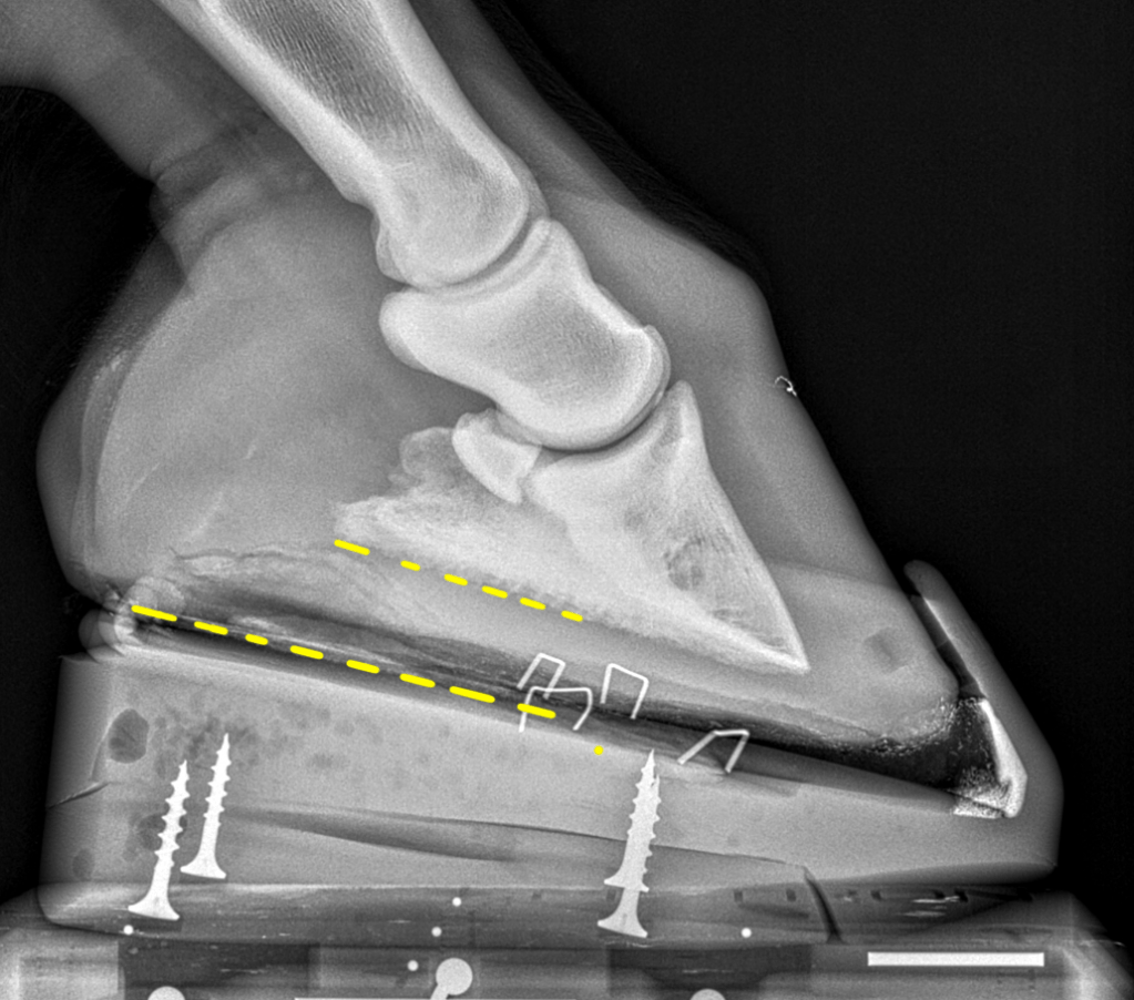
The image at the far right includes the Ultimate which has been attached to the hoof with the bottom of the coffin bone parallel to the surface of the Ultimate. This is the most important thing because it shifts the horse’s weight to the heels, away from the toe. If we applied the Ultimate without getting the coffin bone parallel to the cuff then we would be increasing pressure at the toe making things worse.
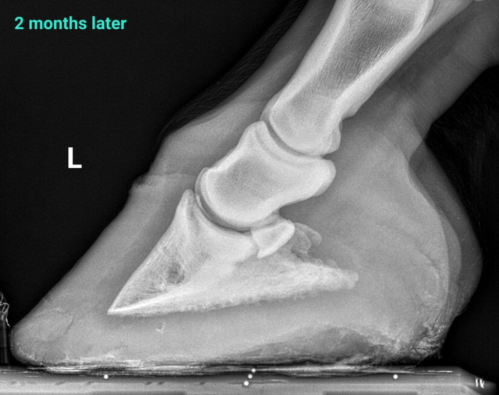
2 months later the horse is sound, he has doubled his sole depth, and improved alignment. I gradually reduced the size of heel elevation and finally removed the Ultimates.
Canker
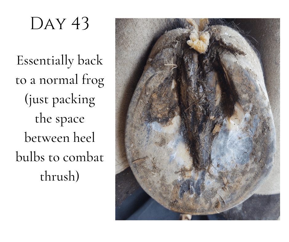
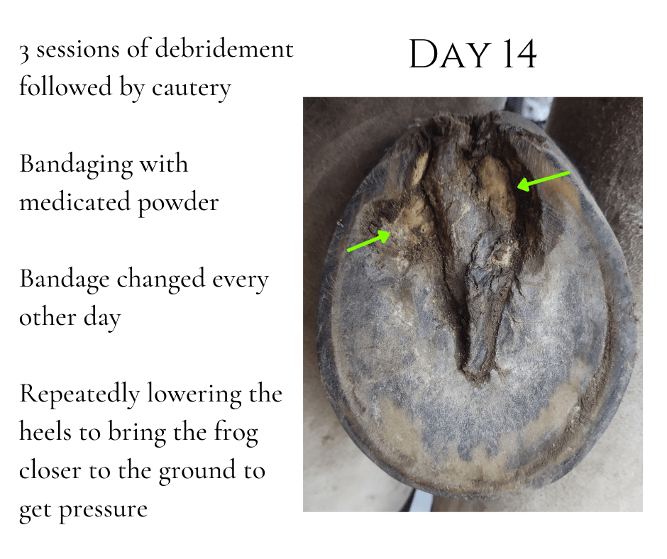
30 year old Tennessee Walking Horse gelding with canker in 3 of 4 hooves
“Canker” is believed to be an anaerobic infection of the superficial epithelium of the hoof. Typically, it affects the frog, bars, and/or sole. The infection is characterized by abnormal keratin proliferation – it has a “wet cauliflower” appearance. The abnormal tissue is often extremely sensitive and bleeds easily.

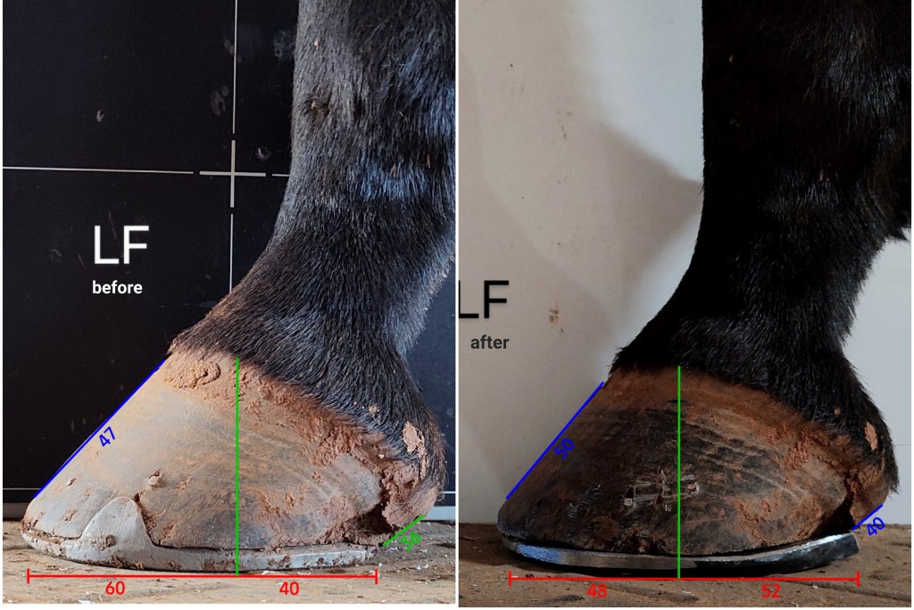
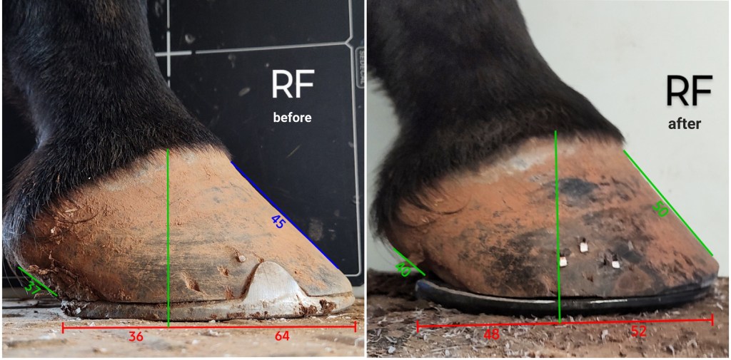
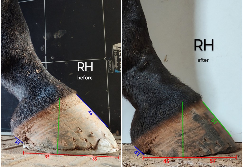
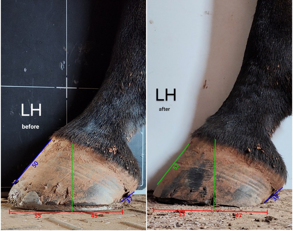
Normal Horse 8 weeks after last shoeing
This horse was very sore over her gluteal muscle palpation which I attributed to her long hind toes/breakover distances (see research on this subject: link 1, link 2). She had been shod about 8 weeks prior to my exam. Measurements were made using HoofmApp. The angles are in green and blue. Ideally the heel angle and the dorsal hoof wall angle should be within about 10 degrees of each other. The red numbers indicate the percentage of hoof in front of the center of rotation (green line) and the percentage behind the center of rotation (CoR). I typically aim for this ratio to be about 50:50.